Image

Image caption
Fig. 4.18. Common orientation terms applied to three different animals: a billfish, a horse, and a person.
Image copyright and source
Image by Byron Inouye
Anatomy is the study of an organismвҖҷs structures. Fishes come in a diverse array of forms, many with special modifications. The shape, size, and structure of body parts permit different fishes to live in different environments or in different parts of the same environment. The external anatomy of a fish can reveal a great deal about where and how it lives.
When describing the basic anatomy of an organism, it is useful to have some common terms to help with orientation. Just as a map uses north, south, east, or west to help determine the location, orientation words are useful in describing anatomy. Table 4.3 defines common anatomy terms, Fig. 4.18 shows their orientation on three different animals.
Fig. 4.18. Common orientation terms applied to three different animals: a billfish, a horse, and a person.
Image by Byron Inouye
| Anatomy Word | ...of the organism |
|---|---|
| Anterior | The head end... |
| Posterior | The tail end... |
| Dorsal | The back |
| Ventral | The front or belly |
| Lateral | The side or flank |
Scientists measure and describe the external features of fishes to identify species, assess age and health, and learn about structure and function. Scientists work with a variety of types of fishes to do this. They might use a fresh fish, or they may use photographs, scientific drawings, or other kinds of detailed imagesвҖ“even fish fossils.

Fig. 4.19. A gyotaku print of a longtooth grouper
Image courtesy of J.G. Wang, Flickr
One way to document details about a fish is gyotaku. Gyotaku (pronounced gee yo TAH koo) is a traditional Japanese method of printmaking, which uses the whole fish. This method can produce an accurate image of a fish (Fig. 4.19).
Gyotaku is a relatively new art form that developed in Japan, probably in the early- to mid-nineteenth century. Gyotaku means вҖҳfish rubbing.вҖҷ Gyotaku is valued from both a scientific and artistic perspective. The detail captured in gyotaku, especially in historical prints, is an important source of information for scientists who want to know the size and external features of fish in the past. The color and artistic arrangement of gyotaku prints made by skilled artists also make them valuable pieces of art. The oldest known gyotaku print, made in 1862, is owned by the Homma Museum in Sakata, Japan.
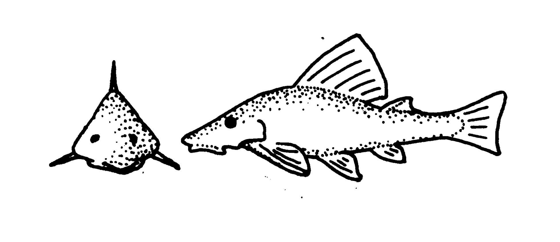
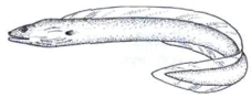
Perches are the most common type of bony fishes. As a result, people often use the words perch-like to describe a generic fish shape. (Fig. 4.21 A). Fusiform is the scientific term used to describe the perchвҖҷs streamlined, torpedo shaped body. Compressiform means laterally flattened (Fig. 4.21 B). Depressiform means dorso-ventrally flattened (Fig. 4.21 C). Anguilliform means eel-like (Fig. 4.21 D). See Table 4.4 for additional descriptions of fish body shapes.
</p>")
Fig. 4.21. (A) Perch (fusiform-torpedo shape)
Image Courtesy of U.S. Department of Agriculture (USDA)
</p>")
Fig. 4.21. (B) Angelfish (compressiform-flat side to side)
Image Courtesy of National Oceanic Atmospheric Administration (NOAA)

Fig. 4.21. (C) Flounder (depressiform-flat top to bottom)
Image courtesy of Katie Samuelson

Fig. 4.21. (D) Eel (anguiliform-eel like)
Image courtesy of Drow male, Wikimedia Commons
Table 4.4. Fish form and function: body shape
| Diagram of Body | Description | Adapted Function |
|---|---|---|
|
Image

|
Anguiliform (eel shape) | Maneuvering in crevasses |
|
Image

|
Fusiform (bullet, or torpedo shape) | Lowering frictional resistance in fast swimmers |
|
Image

|
Depressiform (broad shape and flat top to bottom) | Lying on or below the surface of the sand |
|
Image

|
Compressiform (tall, thin shape and flat side to side) | Entering vertical crevices |
|
Image
|
Vertically flattened shape that is somewhat depressiform (flat top to bottom) | Bottom heavy for sitting on the bottom, not casting a shadow |
|
Image

|
Fusiform (bullet, or torpedo shape), which is also sometimes called perch like | General all-purpose shape |
|
Image
|
Elongated shape that is somewhat anguiliform (eel shape) | Ambush predators |
Images by Byron Inouye

Fig. 4.9. (B) Anatomy of a soldierfish, Myripristis berndti
Image by Byron Inouye
The first anatomical structures many people identify on a fish are the fins. In fact, вҖңappendages, when present, as finsвҖқ is part of one of the scientific definitions of a fish. Most fish have two kinds of fins: median and paired.
Median fins are single fins that run down the midline of the body. The dorsal fin is a median fin located on the dorsal side of the fish. The anal fin and caudal fin are also median fins. Paired fins are arranged in pairs, like human arms and legs. The pelvic and pectoral fins are both paired fins. (Table 4.5).
Table 4.5. Fish form and function: dorsal fin features
| DORSAL FIN DIAGRAM | DESCRIPTION | ADAPTED FUNCTION |
|---|---|---|
|
Image

|
Spiny and soft-rayed dorsal fin. | Flared to make the fish look bigger |
|
Image

|
Tucked dorsal fin | Reduces drag in fast swimming fish |
|
Image

|
Locking spiny dorsal fin | Locking fish into coral crevices |
|
Image

|
Very long dorsal fin | Snake-like locomotion |
|
Image
|
Three dorsal fins | Locomotion |
|
Image
|
No dorsal fin | Snake-like locomotion |
Images by Byron Inouye
Median Fins
Median fins, like the dorsal, anal, and caudal fins, can function like the keel of a boat and aid in stabilization (Fig. 4.22 A). Median fins can also serve other purposes, like protection in the lion fish (Fig. 4.22 B).

Fig. 4.22. (A) The keel of boat stabilizes the boat, similar to a fishвҖҷs anal fin
Image courtesy of Brother Magneto Flickr

Fig. 4.22.(B) The dorsal fin of a lionfish has spines and poison for protection
Image courtesy of Katie Samuelson
Caudal (Tail) Fin
The caudal fin is known commonly as the tail fin (Table 4.6). It is the primary appendage used for locomotion in many fishes. The caudal fin is also a median fin (Fig. 4.22 A).
The caudal peduncle is the base of the caudal fin. Peduncle means stem, and the caudal peduncle is where the strong swimming muscles of the tail are found. Together, the caudal fin acts like a вҖңpropellerвҖқ for the fish, and the caudal peduncle acts like a motor.
Table 4.6. Fish form and function: Caudal fin features
| Tail Diagram | Description | Adapted Function |
|---|---|---|
|
Image

|
Rounded tail | Slow swimming, accelerating, and maneuvering |
|
Image

|
Truncated (triangular) tail | Turning quickly |
|
Image

|
Lunate (moon shaped) tail | Continuous long distance swimming |
|
Image

|
Forked tail | Rapid swimming, somewhat sustained with bursts of speed |
|
Image
|
Heteroceral (taller upper lobe) tail | Slow or rapid swimming with bursts of speed |
Images by Byron Inouye

Fig. 4.25. (A) trout showing two dorsal fins on top and, from left to right, pectoral, pelvic, and anal fins
Photo courtesy of the Wild Center Flickr

Fig 4.25. (B) wrasse
Photo courtesy of Katie Samuelson
Paired Fins
Fish have two sets of paired fins: pectoral and pelvic (Fig 4.25). The pectoral fins are vertical and are located on the sides of the fish, usually just past the operculum (Table 4.7). Pectoral fins are similar to human arms, which are found near the pectoral muscles. Many fish, such as reef fish like wrasses (Fig. 4.25 B), use their pectoral fins for locomotion.
Table 4.7. Fish form and function: Pectoral fin features
| Pectoral Fin Diagram | Description | Adapted Function |
|---|---|---|
|
Image

|
Fringe-like pectoral fins | Probing substrate |
|
Image

|
Spiny pectoral fins | Propping on substrate |
|
Image

|
Hand-like pectoral fins | Crawling on substrate |
|
Image

|
Wing-like pectoral fins | Soaring and swimming |
|
Image
|
No pectoral fins | Snake-like swimming |
|
Image

|
Normal size pectoral fins | Maneuvering |
Images by Byron Inouye
The pelvic fins sit horizontally on the ventral side of the fish, past the pectoral fins (Table 4.8). Pelvic fins are similar to legs. Just like human legs, pelvic fins are associated with the pelvis of the fish.
Table 4.8. Fish form and function: Pelvic Fin Features
| Pelvic Fin Diagram | Description | Adapted Function |
|---|---|---|
|
Image

|
Sucker-like pelvic fins | Grabbing rocks by sucktion |
|
Image

|
Thickened rays on pelvic fins | Sitting on substrate |
|
Image

|
Moderate sized pelvic fins | Locomotion |
Unique and Specialized Fins
Paired fins are most commonly used for maneuvering, like the oars on a rowboat. However, both the pectoral and pelvic fins can also be highly specialized like those of the flying fish (Fig. 4.26 A). Unique combinations of other fins can also help fish to be even more specialized, like the pectoral and anal fins of a box fish (Fig. 4.26 B; see Table 4.9) .

Fig. 4.26. (A) Flying fish with highly specialized pectoral and pelvic fins for flying
Image courtesy of Theron Trowbridge Flickr

Fig. 4.26 (B) Spotted boxfish with specialized dorsal and anal fins for moving its boxy body
Image courtesy of Katie Samuelson
Table 4.9. Fish form and function: Combinations of Fins
| Fin Combination Diagram | Description | Adapted function |
|---|---|---|
|
Image
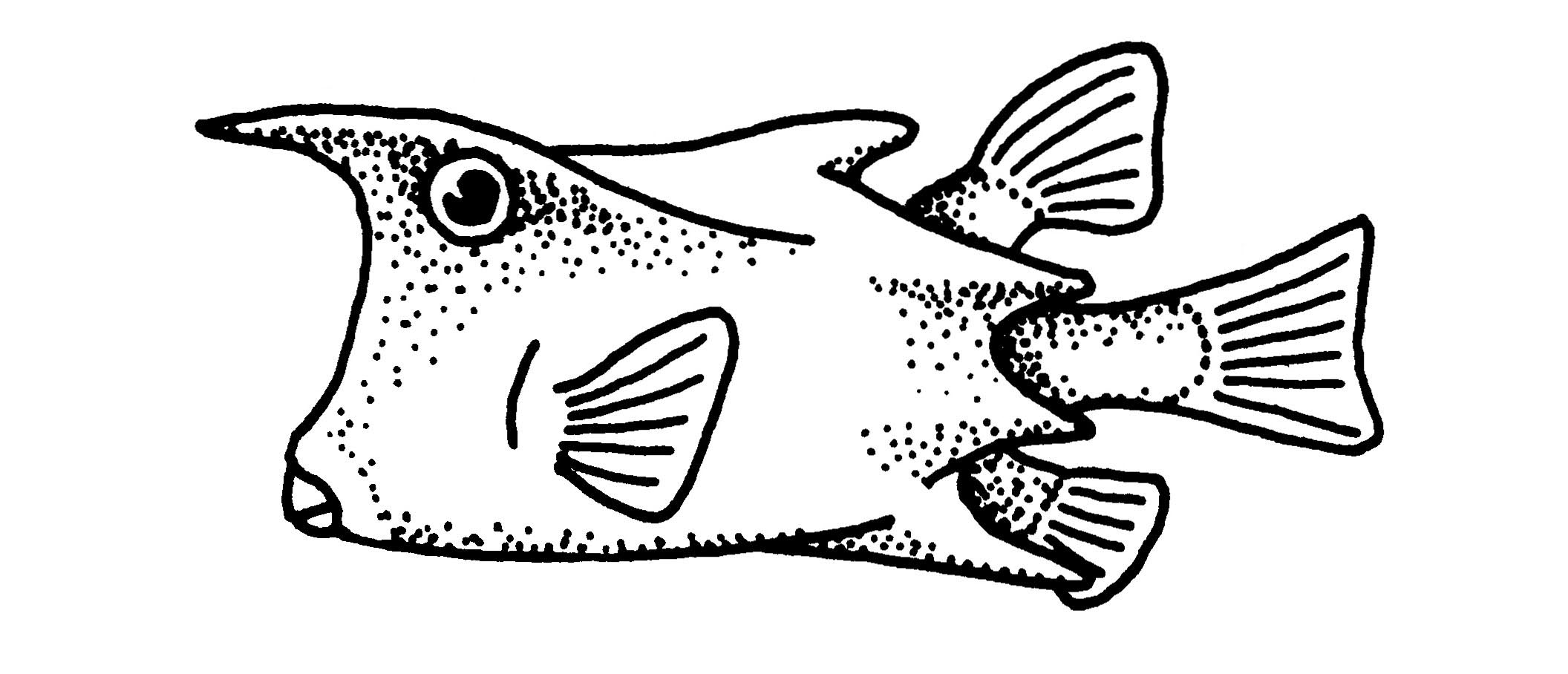
Image copyright and source
Byron Inouye |
Dorsal and anal fins | Modified to increase propulsion |
|
Image

Image copyright and source
Byron Inouye |
Pectoral and tail fins | Modified for soaring in air |
Scientists use fins to help identify and classify fish species. In more evolutionarily advanced fish, the fins are supported by bony structures: spines and soft rays. Spines are simple, unbranched, structures. Soft rays are compound, segmented, and branched structures (Fig. 4.27).

Fig. 4.27. (A) The elongated dorsal fin of a common carp, with 1 spine and 15-22 soft rays.
Image courtesy of John Lyons, University of Wisconsin Sea Grant http://www.seagrant.wisc.edu/home/Default.aspx?tabid=605&FishID=33

(B) A dorsal fin drawing of a soldierfishвҖҷs second dorsal fin, showing fin spines (unbranched) and rays (branched and softer than spines).
Image from Living Ocean, CRDG, University of Hawaii at Manoa
The mouth is at the front, or anterior end, of the fish. The mouth can reveal a lot about the fishвҖҷs feeding habits (Table 4.10). The size, shape, and placement of the mouth, combined with the type of teeth, provide critical information about the feeding habits of a fish (Table 4.11).
For example, a fish with a mouth on the bottom of its head often feeds by digging in the bottom sediment (Fig. 4.28 A). A fish with a mouth oriented upward usually feeds in the water column, or even above the water (Fig. 4.28 B). When a fish has its mouth open, the front lip may slide down and out from the mouth. This sliding action of the mouth can help the fish create a vacuum and quickly suck in a big mouthful of water, which hopefully also includes prey!
Fig. 4.28. (A) A bottom facing mouth indicates bottom feeding preferences in the sturgeon. (B) An upward facing mouth shows the surface feeding adaptation of the arowana.
Table 4.10. Fish form and function: Mouth Features
| Mouth Diagram | Description | Adapted Function |
|---|---|---|
|
Image

Image copyright and source
Byron Inouye |
Jawless | Scavenging or parasitic behavior |
|
Image

Image copyright and source
Byron Inouye |
Tweezer-like snout | Poking into crevices |
|
Image

Image copyright and source
Byron Inouye |
Suction tube | Slurping in prey |
|
Image

Image copyright and source
Byron Inouye |
Large mouth | Swallowing large prey |
|
Image

Image copyright and source
Byron Inouye |
Beak-like teeth | Biting hard objects |
|
Image

Image copyright and source
Byron Inouye |
Tiny and turned up | Capturing plankton |
Table 4.11. Fish form and function: Teeth Features
| Teeth Diagram | Description | Adapted Function |
|---|---|---|
|
Image

Image copyright and source
Byron Inoyue |
Pointed | Stabbing |
|
Image

Image copyright and source
Byron Inouye |
Comb-like | Scraping material off rocks |
|
Image

Image copyright and source
Byron Inouye |
Heavy and flat, molar like | Grinding |
|
Image

Image copyright and source
Byron Inouye |
Fused like a beak | Scraping hard materials off rocks |
|
Image
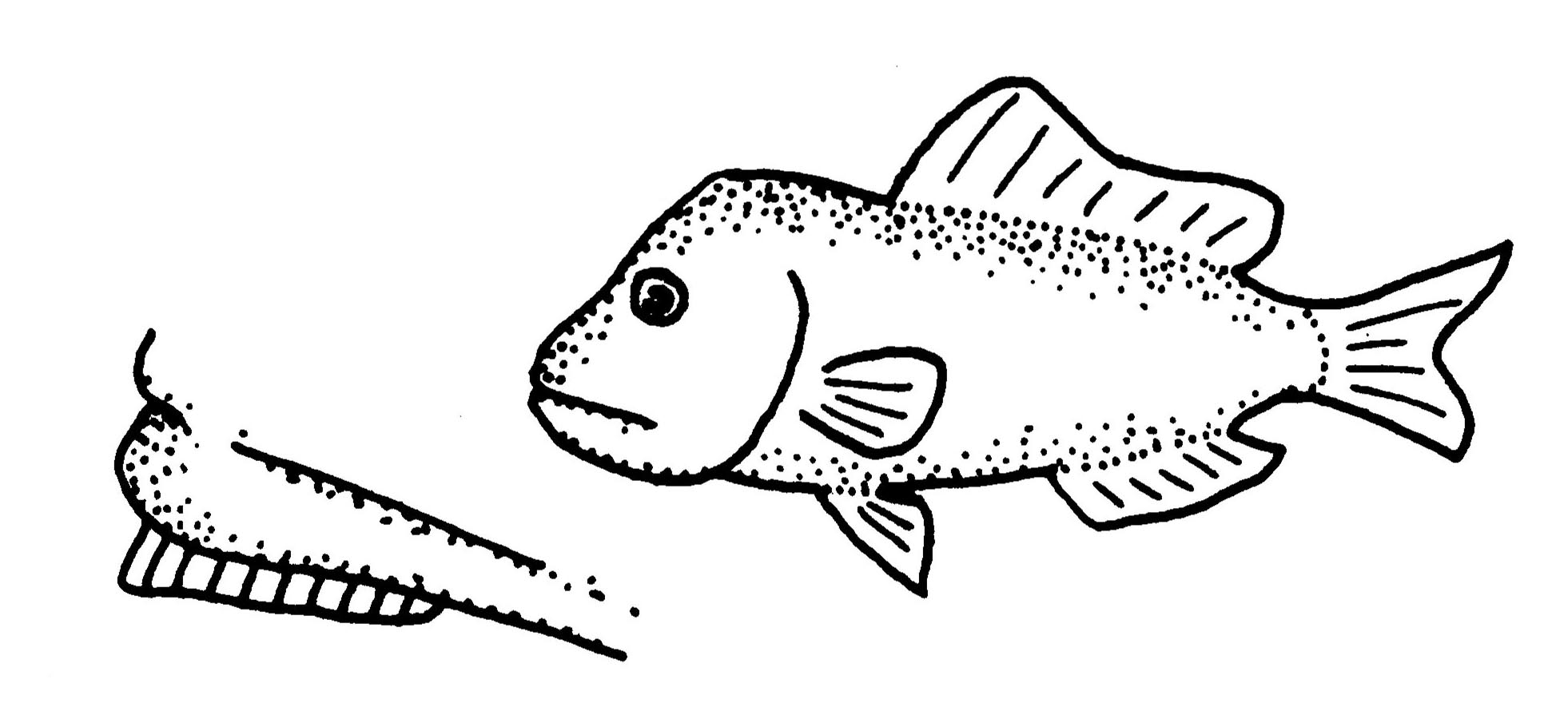
Image copyright and source
Byron Inouye |
Incisor-like | Cutting |
|
Image
Image copyright and source
Byron Inouye |
Broom-like | Filtering |
| Steak knife-like | Serrated for sawing |
The eyes of fish resemble human eyes (Fig. 4.29). At the front of each eye is a lens, held in place by a suspensory ligament. The lens focuses images of objects on the retina. To bring near and far objects into focus, the lens retractor muscle moves the lens back and forth.

Fig. 4.29 Eye of a bigeyed sixgill shark (Hexanchus nakamurai)
https://en.wikipedia.org/wiki/File:Hexanchus_nakamurai_JNC2615_Eye.JPG
Images courtesy of Jean-Lou Justine
The retina is a light-sensitive membrane rich in nerves that connect to the optic lobes of the brain by optic nerves. When light shines on the nerves of the retina, the optic nerves send impulses to the optic lobes. Because fish have no eyelids, their eyes are always open.
Some elasmobranchs, and most teleost fishes, have color vision. Some fishes can also see in ultraviolet (UV) light. UV vision is especially useful for reef fishes. UV vision helps fishes in foraging, communication, and mate selection.
Elasmobranchs, and some teleosts, also have a tapetum lucidum. The tapetum lucidum is a shiny, reflective structure that reflects light and helps vision in low light situations. The tapetum lucidum is what makes the eyes of sharks and deep sea fish, as well as land mammals like cats and cows, shine at night.
Fish eyes are usually placed just dorsal of and above the mouth. Just like the mouth of a fish, the size, shape, and position of the eyes can provide information about where a fish lives and what it feeds on. For example, fish predators often have eyes facing forward in order to provide better depth perception. Prey fish, on the other hand, often have eyes on the sides of their bodies. This gives them a larger field of view for avoiding predators. (Table 4.12).
Table 4.12. Fish form and function: Eye Features
| Eye Diagram | Description | Adapted Function |
|---|---|---|
|
Image

Image copyright and source
Byron Inouye |
Tiny eyes, head length approximately six times longer than eye width | Receiving high intensity light |
|
Image

Image copyright and source
Byron Inouye |
Large eyes, head length approximately three times longer than eye width | Receiving low intensity light or spotting predators |
|
Image
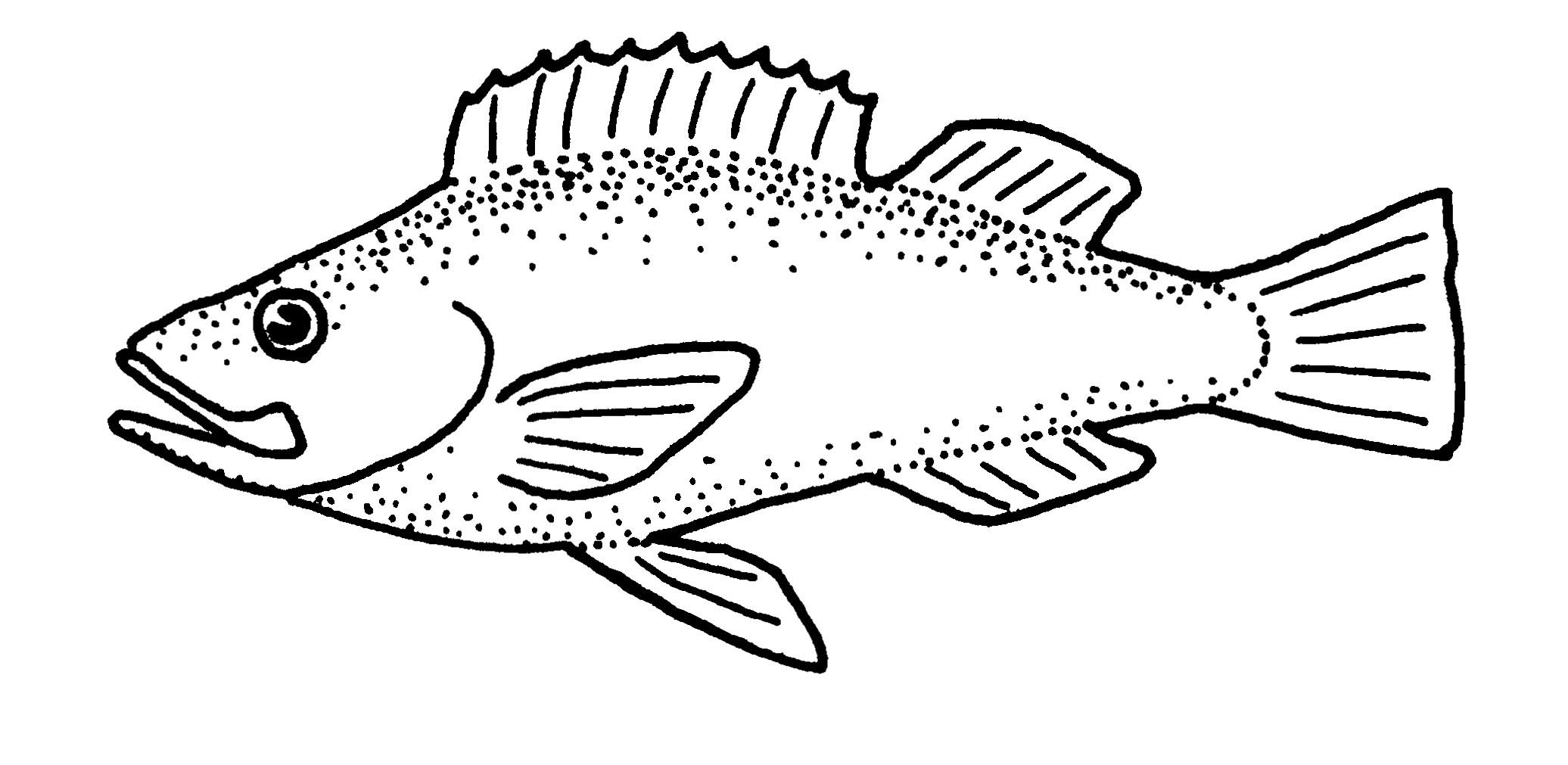
Image copyright and source
Byron Inouye |
Average eyes head length three to five times longer than eye width | Receiving normal intensity light |
|
Image

Image copyright and source
Byron Inouye |
Tubular eyes | Receiving low light from above often in deep water |
|
Image

Image copyright and source
Byron Inouye |
Eyes on dorsal side of the fish | Seeing above |
The sense of smell is well developed in some fishes. Water circulates through openings in the head called nostrils. Unlike humans, fish nostrils are not connected to any air passages. Fish nostrils serve no role in respiration. They are completely sensory.
The largest part of a fishвҖҷs brain is the olfactory lobe, which is responsible for the sense of smell. Smell is the response to chemical molecules by nerve endings in the nostrils. Chemoreception is the scientific term for what nerve cells do to help an organism smell (see Table 4.13).
Taste Receptors
Taste is another form of chemoreception. Fish can taste inside their mouth. Many fishes, like goatfish and catfish, also have fleshy structures called barbels around the chin, mouth, and nostrils (see Table 4.13 and Fig. 4.30). In some fishes, these barbels are used for touch and chemoreception.
Fig. 4.30.

Fig. 4.30. (A) Goatfish with chemosensory barbels that can taste and smell
Image courtesy of Kaite Samuelson

Fig 4.30. (B) Catfish with non-chemosensory barbels, which cannot taste or smell
Photo courtesy of Pil http://www.flickr.com/photos/jp_math54/6130292385/

Fig 4.30. (C) Blenny with non-chemosensory cirri, which cannot taste or smell
Photo courtesy of Katie Samuelson
Not all barbels have chemoreception. The barbels of some fish, like catfishes, are not equipped for chemical reception (Fig. 4.30 B). Some fish also have fleshy tabs called cirri on the head (Fig. 4.30 C). Cirri are not sensory organs.
Table 4.13. Fish form and function: Chemosensory Adaptation and Camouflage
| Diagram | Description | Adapted function |
|---|---|---|
|
Image

Image copyright and source
Byron Inouye |
Barbels | Probing for food in sand. Can detect chemicals for smelling and tasting (but note that not all fishesвҖҷ barbels can detect chemicalsвҖ”like catfish barbels are cannot taste or smell) |
|
Image

Image copyright and source
Byron Inouye |
Tubular nostrils | Detecting chemicals for smelling and tasting |
|
Image
Image copyright and source
|
Cirri on head by eyes | Camouflage (although they resemble chemosensory organs, they do not respond to chemicals) |
Lateral line
Most fish have a structure called the lateral line that runs the length of the bodyвҖ”from just behind the head to the caudal peduncle (Fig. 4.31). The lateral line is used to help fishes sense vibrations in the water. Vibrations can come from prey, predators, other fishes in a school, or environmental obstacles.
Fig. 4.31.

(A) Location of the lateral line on a shark
Image courtesy of Byron Inouye
</p>")
(B) Location of the lateral line on a fish and englarged view of a lateral line, showing the lateral line tube reaching through pores in the fish scales
Image from Living Ocean, CRDG, University of Hawaii at Manoa (Felo 7.17)
The lateral line is actually a row of small pits that contain special sensory hair cells (Fig. 4.32). These hair cells move in response to motion near the fish. The lateral line sense is useful in hunting prey, escaping predators, and schooling.
Fig. 4.32.

Lateral line, close-up of pits with hair cells
Image courtesy of Thomas Haslwanter, Wikimedia Commons https://commons.wikimedia.org/wiki/File:LateralLine_Organ.jpg
Ampullary receptors are sense organs made of jelly-filled pores that detect electricity. They can detect low frequency alternating current (AC) and direct current (DC). Ampullae detect electricity emitted by prey as well as the small electrical fields generated by a fishвҖҷs own movement through the earthвҖҷs magnetic fields. Researchers think that this may help fishes use the EarthвҖҷs magnetic field for navigation. Fishes that have ampullae include sharks, sturgeon, lungfish, and elephant fish. The ampullae of sharks are known as Ampullae of LorenziniвҖ”named for Stefano Lorenzini, who first described them in 1678 (Fig. 4.33).

(A) Ampullae of Lorenzini in a sharkвҖҷs head
Image courtesy of Chris Huh https://en.wikipedia.org/wiki/File:Electroreceptors_in_a_sharks_head.svg

(B) Ampullae of Lorenzini pores on the snout of a tiger shark
Image courtesy of Albert Kok https://en.wikipedia.org/wiki/File:Lorenzini_pores_on_snout_of_tiger_shвҖҰ
Fig 4.33. (A) Ampullae of Lorenzini in a sharkвҖҷs head (B) Ampullae of Lorenzini pores on the snout of a tiger shark
Some fishes can also generate their own electrical fields. These fishes have both ampullae type receptors and tuberous type receptors. The tuberous receptors are most sensitive to the electric organ discharge of the fish itself, which is important for object detection. The tuberous type of receptor is usually deeper in the skin than ampullae.
Some fishes that produce electricity also use it for communication. Electric fishes communicate by generating an electric field that another fish can detect. For example, elephant fishes use electrical communication for identification, warning, submission, courtship, and schooling (Fig. 4.34).

Fig 4.34 The elephant fish use electric impulses to communicate.
Fig 4.34. The elephant fish use electric impulses to communicate.
Sound travels well underwater, and hearing is important to most fishes. Fishes have two inner ears embedded in spaces in their skulls. The lower chambers, the sacculus and the lagena, detect sound vibrations. (See Fig. 4.35.)
</p>")
Fig 4.35. Inner ear of a fish
Image from Living Ocean, CRDG, University of Hawaii at Manoa (Felo 7.16)
Each ear chamber contains an otolith and is lined with sensory hairs. Otoliths are small, stone-like bones (See Fig. 4.36). They float in the fluid that fills the ear chambers. Otoliths lightly touch the sensory hair cells, which are sensitive to sound and movement.

Fig. 4.36. (A) Otolith (ear bone) of an American barrelfishImage courtesy of NOAA Ocean Explorer
Image courtesy of NOAA Ocean Explorer

Fig. 4.36. (B) A pair of otoliths from a 160lb eight-banded grouper
Image by Kanesa Duncan Seraphin
Fig. 4.36. (A) Otolith (ear bone) of an American barrelfish (B) A pair of otoliths from a 160lb eight-banded grouper
Like the otoliths in human ears, otoliths in fishes help with hearing and with balance. When a fish changes position, the otoliths bump the hair cells in the ampullae. The ampullae are bulges in the semicircular canals of the ears (Fig 4.36). When a fish rolls right or left, tail up or tail down, the liquids and otoliths push against the hairlike nerve endings lining the canal, sending messages to the fishвҖҷs brain.
Some fishes also use other organs to aid in hearing. For example, the gas bladder changes volume in response to sound waves. Some fishes can detect these changes in gas bladder volume and use them to interpret sounds.
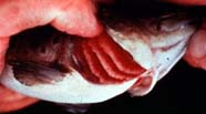
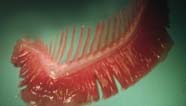
Most mammals get oxygen from the air, but most fishes get oxygen from the water. To get oxygen from the water, fish must pass water over their gills. Gills are composed of a gill arch, gill filaments, and gill rakers (see Fig. 4.37). In many fishes the gill arch is a hard structure that supports the gill filaments. The gill filaments are soft with lots of blood vessels to absorb oxygen from the water.
</a></p>")
Fig. 4.37. (A) A bony fish with the operculum held open to show the gills
Image Courtesy of National Oceanic and Atmospheric Administratrion (NOAA)
</a></p>")
(B) A single gill removed from a bony fish
Image courtesy of National Oceanic and Atmospheric Administration (NOAA)

(C) A drawing of a gill showing gill filaments (oxygen absorption), gill arch (supporting structure), and gill rakers (comb like structure for filtering)
Image from Living Ocean, CRDG, University of Hawaii at Manoa
Fig. 4.37. (A) A bony fish with the operculum held open to show the gills (B) A single gill removed from a bony fish (C) A drawing of a gill showing gill filaments (oxygen absorption), gill arch (supporting structure), and gill rakers (comb like structure for filtering).
As water passes through a fishвҖҷs mouth, over the gills, and back into the environment, oxygen and carbon dioxide are exchanged. Some fishes, like tunas, need to continuously swim to get oxygen from the water. Other fishes, like wrasses, can pass water over their gills by pumping it. This enables wrasses to remain motionless and still get oxygen.
Fishes get both oxygen and food from water. To get oxygen, water needs to move toward the gills. But, to get energy from food, the food needs to move down into the fishвҖҷs stomach. The gill rakers are comb-like structures that filter food from the water before it heads to the gills. This keeps food particles inside the fishвҖҷs mouth and lets water move out toward the gills.
The structure of a fishвҖҷs gill rakers indicates something about its diet. Fish that eat small prey like plankton tend to have many long, thin gill rakers to filter very small prey from the water as it passes from the mouth to the gills. On the other hand, fish that eat large prey tend to have more widely spaced gill rakers, because the gill rakers do not need to catch tiny particles.
The Operculum is the bony plate that covers fishesвҖҷ gills. In chimeras and bony fishes, the operculum covers the posterior end of the head, which protects the gill openings. The bony operculum often has another bony flap, called the preoperculum, overlaying it (Fig. 4.30). Some fishes also have a strong spine, or spines, that project back from the preoperculum or operculum. These spines are usually used for protection.
Sharks and rays have open, naked gills (see Table 4.14), meaning that they are not covered by an operculum. Their classification name, elasmobranch, actually means naked gill. Most elasmobranchs have five gill openingsвҖ”exceptions include the six gill and seven gill shark.
Table 4.14. Fish form and function: Gills
| GILL DIAGRAM | DESCRIPTION | ADAPTED FUNCTION |
|---|---|---|
|
Image

Image copyright and source
Byron Inoyue |
Elasmobranchs have naked gills | Easy water flow |
|
Image

Image copyright and source
Byron Inouye |
Operculum covers gills | Gill protection |
|
Image

Image copyright and source
Byron Inouye |
Preoperculum and operculum spines | Armor and protection |

Fig. 4.38. (A) A semicircle angelfish (Pomacanthus semicirculatus) with bright blue highlight color on the preoperculum, preoperculum spine, and operculum
Image by Stan Shebs

(B) A dog snapper (Neomaenis jocu) with preoperculum, operculum, and operculum spine labeled.
Image by Byron Inouye
Fig. 4.38. (A) A semicircle angelfish (Pomacanthus semicirculatus) with bright blue highlight color on the preoperculum, preoperculum spine, and operculum (B) A dog snapper (Neomaenis jocu) with preoperculum, operculum, and operculum spine labeled.
The buccal pump is what fish use to move water over their gills when they are not swimming. The buccal pump has two parts: the mouth and the operculum. During the first stage of pumping, both opercula close, and the mouth opens. Water then enters through the mouth. Next, the fish closes its mouth and opens its opercula so that water moves over the gills, which remove oxygen from the water. Some fishes also use the buccal pump as part of their feeding strategy by filtering out small organisms living in the water as they pump water (Fig. 4.39). As water passes through, the gill rakers help to trap plankton from the water.

Fig. 4.39. Some fishes feed by filtering out through their buccal pump such as this whale shark, which feeds on plankton
Image by Arturo de Frias Marques https://en.wikipedia.org/wiki/File:Whale_Shark_1_AdF.jpg
Fig. 4.39. Some fishes feed by filtering out through their buccal pump such as this whale shark, which feeds on plankton
A pore is a small opening in the skin. A typical fish has anal, genital, and urinary pores located anterior of the anal fin. The anal pore is where feces exits the fish body. The anus is the largest and most anterior of the pores (Fig. 4.40 A).
The genital pore is where eggs or sperm are released. The urinary pore is where urine exits the body. Often the genital and urinary pore are combined into a single urogenital pore. These pores are situated on a small papilla, or bump, just behind the anus (Fig. 4.40 B).

Fig. 4.40. Anal pore (toward the head) and genital and/or urinary pores (toward the tail)
Most fishes reproduce externally, meaning that the sperm and eggs meet outside their bodies. However, some fishes reproduce internally. The females of these fishes often have a genital pore that is modified for internal fertilization.
One definition of a fish includes вҖңbody usually covered with scales.вҖқ Except for some parts of the head and fins, the bodies of many fishes are covered with overlapping scales (Fig. 4.41). Scales generally serve to protect the fishвҖҷs skin.

Fig. 4.41. The overlapping scales of a roach fish (Rutilus rutilus)
Image by kallerna
Different fishes have different types of scales. These different types of scales are made of different types of tissue (Fig. 4.42 and Table 4.15). Types of scales also correspond to evolutionary relationships (Fig. 4.9).
Placoid scales are found in the sharks and rays (Fig. 4.42 A). Placoid scales are made of a flattened base with a spine protruding towards the rear of the fish. These scales are often called dermal denticles because they are made from dentin and enamel, which is similar to the material teeth are made of.
Ganoid scales are flat and do not overlap very much on the body of the fish (Fig. 4.42 B). They are found on gars and paddlefishes. In the sturgeon, ganoid scales are modified into body plates called scutes.
Cycloid and Ctenoid scales are found in the vast majority of bony fishes (Figs 4.42 C and 4.42 D). These types of scales can overlap like shingles on a roof, which gives more flexibility to the fish. These scales also form growth rings like trees that can be used for determining age.
Ctenoid scales are different than cycloid scales in that cycloid scales tend to be more oval in shape. Ctenoid scales are more clam shaped and have spines over one edge. Cycloid scales are found on fishes such as eels, goldfish, and trout. Ctenoid scales are found on fishes like perches, wrasses, and parrotfish. Some flatfishes, like flounder, have both cycloid and ctenoid scales.

Fig. 4.42. Four types of fish scales A) Placoid, B) Ganoid, C) Cycloid, and D) Ctenoid
Image from Living Ocean, CRDG, University of Hawaii at Manoa
Table 4.15. Fish form and function: Scale Features
| Scale Diagram | Description | Adapted Function |
|---|---|---|
|
Image

Image copyright and source
Image from Living Ocean, CRDG, University of Hawaii at Manoa |
Spines | Protection from predators |
|
Image
Image copyright and source
Image from Living Ocean, CRDG, University of Hawaii at Manoa |
Blades | Protection and defense |
|
Image

Image copyright and source
Image from Living Ocean, CRDG, University of Hawaii at Manoa |
Scutes (or keel; not shown) | Cuts through water, streamlines swimming |
|
Image

Image copyright and source
Image from Living Ocean, CRDG, University of Hawaii at Manoa |
Many large scales | Protection |
|
Image

Image copyright and source
Image from Living Ocean, CRDG, University of Hawaii at Manoa |
No scales | Burrowing |
|
Image

Image copyright and source
Image from Living Ocean, CRDG, University of Hawaii at Manoa |
Leathery scales | Protection |
|
Image

Image copyright and source
Image from Living Ocean, CRDG, University of Hawaii at Manoa |
Bony armor scutes | Protection from predators |
|
Image

Image copyright and source
Image from Living Ocean, CRDG, University of Hawaii at Manoa |
Rough scales | Protection from parasites and swimming locomotion |
|
Image

Image copyright and source
Image from Living Ocean, CRDG, University of Hawaii at Manoa |
Regular scales | Protection |

Fig. 4.43. (A) Pinecone fish
Photo courtesy of Spencer77 http://www.flickr.com/photos/spencer77/4804550258/

(B) porcupine fish
Photo courtesy of Katie Samuelson
Scale size varies greatly among species, and not all fishes have scales. Some fishes, like some rays, eels, and blennies, do not have any scales. This is probably because these fishes spend a lot of time rubbing on the sand or in rocks. If they had scales, the scales would likely rub off. At the other extreme, some fishes have scales modified into bony plates, such as on a sturgeon and pinecone fish (Fig. 4.43 A). Other fish have scales modified into spines for protection, like the porcupine fish (Fig. 4.43 B).
Fishes are very diverse, and there are examples of extreme body modifications in many different groups of fishes (see Table 4.16). For example, some fishes, like angler fish, have lures to attract prey. Others, like lionfish, have poison sacs to protect them from predators.
Table 4.16. Fish form and function: Other Modifications
| Diagram | Description | Adapted Function |
|---|---|---|
|
Image
Image copyright and source
Image from Living Ocean, CRDG, University of Hawaii at Manoa |
Lures | Attracting prey |
|
Image

Image copyright and source
Image from Living Ocean, CRDG, University of Hawaii at Manoa |
Poison sacs at base of spines | Protection |
Color
The color of fishes is very diverse and depends on where a fish lives. Color can be used as camouflage. Color also plays a role in finding mates, in advertising services like cleaning, in attracting prey, and in warning other fishes of danger (see Table 4.17).
Tunas, barracuda, sharks, and other fishes that live in the open ocean are often silvery or deep blue in color. These fishes also have a body coloring pattern called counter shading. Counter shading means dark on the dorsal, or top, surface and light on the ventral, or belly side. Countershading helps to camouflage fishes by matching the dark, deep water when viewed from above and matching the light, surface water, when viewed from below (Fig. 4.44 B).

Fig. 4.44. (A) blue silvery color in HellerвҖҷs barracuda
Image courtesy of Katie Samuelson

(B) Countershading in a grey reef shark
Image courtesy of Fbattail
Fig. 4.44. (A) blue silvery color in HellerвҖҷs barracuda (B) Countershading in a grey reef shark
Nearer to shore, many fishes have also evolved to be camouflaged in their environment. Kelpfish have developed both colors and a body shape that helps them blend in with the seaweed that they live in. Reef fish often look like coral. Fishes that hide in the sand, like blennies, flat fish, and flounder, are often a speckled sandy color (Fig. 4.45 B).

Fig. 4.45. (A) A leafy seadragon hiding in kelp
Image courtesy of EyeKarma
.jpg\" href=\"https://en.wikipedia.org/wiki/File:Salarias_sinuosus_(Fringelip_blenny).jpg\">Jason Marks</a></p>")
(B) A blenny hiding in coral
Image courtesy of Jason Marks

(C) A three-spot flounder hiding in sand
Image courtesy of Katie Samuelson
Many brightly colored fishes that live in coral reef habitats also use their color, stripes, and spots as camouflage (Fig. 4.46). This is partly because wavelengths of light, and therefore color, appear different under water and change with depth and water color. Water absorbs light. Thus, the amount of light decreases with increasing depth.
Red color, for example, fades out very fast with increasing depth. Fishes with red color, like soldierfish (Fig. 4.46 A), are actually invisible at night and in deep waters. Yellow and blue colors, on the other hand, blend in with the reef color, also providing camouflage from predators (Fig. 4.46 B). Even stripes and spots can prevent an individual fish from standing out, making it harder for a predator to strike (Fig. 4.46 C).

Fig. 4.46. (A) Soldierfish
Image courtesy of Katie Samuelson

(B) blue and yellow Hawaiian cleaner wrasse
Image courtesy of Katie Samuelson

(C) school of convict tang and whitebar surgeonfish
Image courtesy of Katie Samuelson
Fig. 4.46. (A) Soldierfish (B) blue and yellow Hawaiian cleaner wrasse (C) school of convict tang and whitebar surgeonfish
In addition to colors visible to humans, fish also use ultraviolet (UV) light colors for camouflage and communication. Some fishes can see using UV light, and so they use UV colors to identify each other and to avoid predators. Many reef fish can also blink their colors on and off to flash messages (Fig. 4.47). Skin cells called chromatophores allow fish and other animals to quickly change skin color.

Fig. 4.47. Examples of color-changing fish. The peacock flounder (Bothus mancus or pДҒkiвҖҳi in Hawaiian) is a bottom-dwelling flatfish common in the tropical Pacific. It can rapidly change skin colors.
Image courtesy of Brocken Inaglory, Wikimedia Commons
Table 4.17
| Body Color Diagram Example | Picture Example | Description | Adapted Function |
|---|---|---|---|
|
Image

Image copyright and source
Image from Living Ocean, CRDG, University of Hawaii at Manoa |
Image

Image copyright and source
Image Courtesy of Wikimedia Commons |
Both sexes brightly colored | A warningвҖ”not good to eat |
|
Image

Image copyright and source
Image from Living Ocean, CRDG, University of Hawaii at Manoa |
Image

Image copyright and source
Image Courtesy of Wikimedia Commons |
Brightly colored areas around the spine on the caudal peduncle area. The spine is used in defense. | Warning |
|
Image

Image copyright and source
Image from Living Ocean, CRDG, University of Hawaii at Manoa |
Image

Image copyright and source
Image Courtesy of Wikimedia Commons |
Mottled | Camouflage |
|
Image

Image copyright and source
Image Courtesy of Living Ocean, University of Hawaii at Manoa |
Image

Image copyright and source
Image Courtesy of Hans-Petter Fjeld |
Dark on top, lighter on bottom | Camouflage in midwater |
|
Image

Image copyright and source
Image from Living Ocean, CRDG, University of Hawaii at Manoa |
Image

Image copyright and source
Image Courtesy of Wikimedia Commons |
Dark all over | Camouflage in dark areas |
|
Image

Image copyright and source
Image from Living Ocean, CRDG, University of Hawaii at Manoa |
Image

Image copyright and source
Image Courtesy of Wikimedia Commons |
Red all over | Camouflage in dark areas |
|
Image

Image copyright and source
Image from Living Ocean, CRDG, University of Hawaii at Manoa |
Image

Image copyright and source
Image Courtsey of Wikimedia Commons |
Light all over | Camouflage in light areas |
|
Image

Image copyright and source
Image from Living Ocean, CRDG, University of Hawaii at Manoa |
Image

Image copyright and source
Image Courtesy of Wikimedia Commons |
Eyespots | Leading predator away from head |
|
Image

Image copyright and source
Image from Living Ocean, CRDG, University of Hawaii at Manoa |
Image

Image copyright and source
Image Courtesy of Wikimedia Commons |
Brightly colored | Camouflage or communication |
Living things are composed of cells. Cells often become specialized to perform certain functions. For example, muscle cells contract, nerve cells transmit impulses, and gland cells produce chemicals. A tissue is a group of similar cells performing a similar function (Fig. 4.48). There are many kinds of tissuesвҖ”bone, cartilage, blood, fat, tendon, skin, and scales.

Fig. 4.48. Organization of structures in living organisms
Image from Living Ocean, CRDG, University of Hawaii at Manoa
An organ is a group of different kinds of tissues working together to perform a specific function (Fig. 4.48). The stomach is an example of an organ made of several types of tissues.
вҖў Muscle tissue, in the wall of the stomach, contracts to churn and mix food.
вҖў Glandular tissue, in the inner lining of the stomach, secretes digestive chemicals (enzymes).
вҖў Nerve tissue, in the wall of the stomach, coordinates mixing and digesting activities.
An organ system is a group of organs that together perform a function for the body. The digestive system, for example, consists of organs such as the mouth, the stomach, and the intestine (Fig. 4.48). These organs work together to break down food and provide nutrients to the body.
An organism is an entire living thing with all its organ systems (Fig. 4.48). A complex organism like a fish has digestive, nervous, sensory, reproductive, and many other systems. Fish consist of interacting groups of organ systems that together enable a fish to function.
The integumentary system is commonly called the skin. It consists of two layers, the epidermis, or outer layer, and the dermis, or inner layer. Beneath these are the muscles and other tissues that the skin covers (Fig. 4.49).

Fig. 4.49. (A) The skin and cycloid scales of a rohu fish

(B) A drawing of the skin and integumentary system of a fish, showing scales, epidermis, dermis, and muscle
Image from Living Ocean, CRDG, University of Hawaii at Manoa
The epidermis is the top layer of the integumentary system. It is made of several sheets of cells that cover the scales. As the cells age, new cells growing underneath push older cells toward the outer surface.
In the epidermis of most fishes are cells that produce mucus, a slippery material like runny gelatin, that helps the fish slide through the water. The mucus wears off daily, carrying away microscopic organisms and other irritants that might harm the fish. The odor typical of most fish comes from chemicals in the mucus.
In their epidermis, fishes have cells containing pigment grains that give the fish its color. Some fish can change color by expanding or contracting pigment cells. The changes are controlled by hormones that are produced by the endocrine system and regulated by the nervous system.
The lower layer of the integumentary system contains blood vessels, nerves for sensing touch and vibration, and connective tissue made of strong fibers. A special layer of dermal cells secretes chemicals to produce scales, which grow larger as the fish grows. Most fish have covering scales that protect them from damage when they bump into things or are attacked. As the scales grow, they form concentric rings in some fishes. These growth rings can be used to determine a fishвҖҷs age. A few fish, such as catfish, have no scales.
The skeletal system supports the soft tissues and organs of the fish (Fig. 4.50). The skeleton also protects organs and gives the body of the fish its basic shape. The many bones of the skull form a rigid box that protects the brain. Holes, hinges, and pockets in the skull allow room for the nostrils, mouth, and eyes.

Fig. 4.50. (A) The skeleton of a cod fish
Image courtesy of Olaf

(B) A drawing of a fish skeletal system
Image from Living Ocean, CRDG, University of Hawaii at Manoa
Fig. 4.50. (A) The skeleton of a cod fish (B) A drawing of a fish skeletal system
The vertebral column, or backbone, is not a solid rod. The backbone is actually a string of small bones called vertebrae. See Fig. 4.51. Each vertebra has a small hole in it. Together, the small holes in the vertebrae form a canal through which the spinal cord passes. The vertebrae bones protect the spinal cord. Spaces between the vertebrae allow the backbone to bend and nerves to reach the tissues and organs of the body. Rib bones protect the body cavity. Additional bones support the spines and rays.

Fig. 4.51. (A) A photo of the vertebrae of a small fish
Image Courtesy of Assianir

(B) A drawing of a fish skeleton vertebrae viewed from the front, showing rib and tail sections
Image from Living Ocean, CRDG, University of Hawaii at Manoa
Muscles are tissues that contract to shorten and relax to lengthen. Fish move by contracting and relaxing their muscles. Like humans, fish have three types of muscles: skeletal muscles, smooth muscles, and heart muscles.
The muscles and bones of a fish work together. Skeletal muscles use bones as levers to move the body. Tendons are strong connective tissues that attach muscle to bone. When muscle cells are stimulated, they contract and shorten, which pulls on tendons to move bones.
Skeletal muscles are voluntary, meaning that they move only when the thinking part of the brain signals them to move. To swim, fish must contract and relax their skeletal muscles, just as humans do when they learn to walk. Most of a fishвҖҷs body is made of layers of skeletal muscle. These layers are arranged in W-shaped bands from belly to back (Fig. 4.52). This network of muscles is vertical and interlocking, which allows the fish to move the body back and forth in a smooth, undulating motion. Such motion would not be possible if the muscles ran horizontally along the length of the body, from head to tail.

(A) Side view of salmon skeletal
Image courtesy of Flying Penguin

(B) Drawing of skeletal muscle pattern in a fish
Image from Living Ocean, CRDG, University of Hawaii at Manoa
Fig. 4.52. (A) Side view of salmon skeletal muscle (B) Drawing of skeletal muscle pattern in a fish
A fish swims by alternately contracting muscles on either side of its body (See Fig. 4.53 B). Swimming begins when the muscles on one side of the body contract, pulling the caudal fin toward that side. The sideways movement of the caudal fin pushes the fish forward. Then the muscles on the opposite side of the body contract, and the caudal fin moves toward the other side of the body.

(A) Sardines swim by contracting their tail muscles
Image courtesy of James Kilfiger

(B) A drawing contrasting a typical fish swimming movement with the movement of a typical human swimming with dive fins.
Image from Living Ocean, CRDG, University of Hawaii at Manoa
Fig. 4.53. (A) Sardines swim by contracting their tail muscles (B) A drawing contrasting a typical fish swimming movement with the movement of a typical human swimming with dive fins.
Skeletal muscles are also attached to bones that move the fishвҖҷs paired fins. Fishes with wide pectoral fins, like wrasses, swim by flapping their pectoral fins. Other fishes, like fast-swimming tunas, move mostly with their caudal fin but use long, thin pectoral fins for steering.
Skeletal muscles also move dorsal fins. Faster-swimming fishes reduce water drag by tucking in their dorsal fins while swimming. Slower-swimming reef fishes have larger dorsal fins, which they sometimes flare to protect themselves in encounters with other fish.
Smooth muscles move internal organs of the body and line tubes such as the intestinal tract and blood vessels. Smooth muscles are involuntary; they move without signals from the thinking part of the brain. For example, smooth muscles automatically contract and relax to push food through the digestive tract from the mouth to the anus. Other smooth muscles control the flow of blood and other body fluids and movement in the urogenital tract.
Heart muscles are also involuntary. However, the structure of heart muscle cells is different from involuntary smooth muscles, so these two muscle types are given separate names. Heart muscles pump blood through the blood vessels by rhythmically contracting and relaxing.
Respiratory System
The respiratory system takes oxygen (O2) into the body and passes carbon dioxide (CO2) out of the body. Oxygen is essential to fishвҖҷs digestion because it combines with food molecules to release energy for the fishвҖҷs needs.
The respiratory organs in fish are gills. Each gill has many gill filaments, which contain a network of tiny blood vessels called capillaries (Fig. 4.54). The gill cover (also called the operculum) is the body surface that covers the gills. The gill rakers filter food from the water as water passes out to the gills.
.jpg\" href=\"https://commons.wikimedia.org/wiki/File:Gills_(esox).jpg\">Uwe Gille</a></p>")
Fig. 4.54. (A) Exposed fish gills as viewed from the ventral, or belly side, of the head
Image courtesy of Uwe Gille

(B) A drawing of a gill filament with a gill raker and the gill arch labeled
Image from Living Ocean, CRDG, University of Hawaii at Manoa
Fig. 4.54. (A) Exposed fish gills as viewed from the ventral, or belly side, of the head (B) A drawing of a gill filament with a gill raker and the gill arch labeled
Water moves over the gills in a pumping action with two steps (Fig. 4.55). In the first step, the mouth opens, the gill covers close, and the fish brings water into its mouth. In the second step, the mouth closes, the gill covers open, and water passes out of the fish. This action is called buccal pumping and is named for the cheek muscles that pull water into the mouth and over the gills.
Some fish also use ram ventilation to move water over their gills. When swimming fast, fish like sharks and tunas open both their mouths and gill openings to let water pass continuously through their gills. They do not need to open and close their mouth because water is pushed over their gills by their swimming action.
As water passes over the gills, carbon dioxide in the blood passes into the water through the capillaries of the gill filaments. The same gill filaments allow dissolved oxygen from the water to pass into the blood, which then carries it throughout the body.

Fig. 4.55. Movement of water past the gills
Image from Living Ocean, CRDG, University of Hawaii at Manoa
Fig. 4.55. Movement of water past the gills
Buoyancy refers to whether something will float or sink. Some fishes have a gas bladder that helps control their buoyancy. The gas bladder is a special, gas filled chamber in a fishвҖҷs body cavity. It lies just below the kidneys.
The gas bladder is often called the swim bladder because it regulates buoyancy by making the fishвҖҷs density equal to the density of the surrounding water. The average density of seawater is 1.026 g/mL, but the density of fish flesh and bones is about 1.076 g/mL. This means that a typical fish is denser than seawater and would naturally sink. The density of the gas bladder, on the other hand, is less dense than seawater. The low density of the gas bladder helps the fish float (Fig 4.56).

Fig 4.56. (A) The position of the gas bladder (swim bladder) in a bleak (Alburnoides bipunctatus)
Image courtsey of Wikimedia

(B) Gas bladder from a Ruddy fish (Scardinius erythrophthalmus)
Image Courtesy of Wikipedia
Fig 4.56. (A) The position of the gas bladder (swim bladder) in a bleak (Alburnoides bipunctatus) (B) Gas bladder from a Ruddy fish (Scardinius erythrophthalmus)
The gas bladder has a low density because it is filled mostly with oxygen and nitrogen gases. The gas bladder acts like an inflatable balloon inside the fish. The gas bladder reduces the density of the fishвҖҷs body until it is the same as the density of seawater. This helps the fish float within the water column.
In many groups of fishes (like herring, pike, catfish, eels), an open tube connects the gas bladder to the digestive tract. This allows the fish to adjust gas content in the bladder by swallowing and expelling air through their mouth. Other kinds of fishes (like perches, snappers, groupers) have a gas gland that bubbles gasses into and out of the bloodstream to inflate and deflate the gas bladder.
Pressure increases with increasing water depth because the water above pushes down on the water (and animals) below. When a fish swims into deeper water, its gas bladder gets smaller because of the increase in water pressure. Thus, as a fish goes deeper, it must add gas to its gas bladder to maintain neutral buoyancy. When a fish swims into shallow water, its gas bladder expands because the pressure of water surrounding the fish decreases. Thus, as it moves into shallower water, the fish must absorb gas from the gas bladder to maintain neutral buoyancy.
Because gases move slowly in and out of the gas bladder, fish caught at great depths are often bloated when they are brought to the surface quickly. The gas in the gas bladder expands when the fish moves from the high pressure of deep water to the lower pressure at the surface. A fish pulled quickly to the surface cannot absorb the gases fast enough, and the sudden expansion of the gas bladder can injure the fish (Fig. 4.57).

Fig. 4.57. Inflated gas bladder of a deep water rougheye rockfish after capture.
Image courtesy of NOAA Fisheries: Alaska Fisheries Science Center
To keep the fish alive, collectors must bring fish to the surface slowly to let the fishвҖҷs body absorb the gases from the gas bladder. There are also methods for releasing a fish with recompression in order to help it recover from gas expansion as a result of being brought quickly to the surface (Fig. 4.58).

Fig. 4.58. Recompression is lowering a fish to its natural depth in a controlled manner, using devices like weights and baskets, and allows gas in the gas bladder to be reabsorbed into the fishвҖҷs body.
Image courtesy of NOAA Fisheries: West Coast Region
Some fishes, such as grunts and toadfish, can use their gas bladder to produce sound. Muscles in the wall of the bladder contract rapidly, producing a low-frequency (low-pitch) sound that is resonated and amplified in the bladder. Other fishes, like the lungfish, also use the gas bladder as an accessory respiratory organ or вҖңlungвҖқ when they crawl on land.
Fishes that have no gas bladder are always denser than the surrounding water, so they sink if they stop swimming. Sharks, for example, must keep swimming to stay afloat. They use their tails and pectoral fins like airplane wings, adjusting the amount of lift to control the depth of their swimming. Many bottom-dwelling fishes also lack gas bladders because they have no real need from them.
The circulatory system is a transportation system for body fluids. The circulatory system brings nutrients to cells and carries waste away from cells. Blood is a fluid that consists of plasma (the liquid part) and blood cells. Plasma contains water, carbon dioxide (CO2), hormones, nutrients, wastes, and other molecules. Blood cells are of two main types: red and white.
Red blood cells carry oxygen (O2) from the gills to other cells in the body. In red cells, special molecules that combine chemically with oxygen can pick up and release oxygen, depending on the surrounding environment. These molecules, called hemoglobin, contain iron atoms. When hemoglobin combines with oxygen, it turns bright red. When hemoglobin releases its oxygen, it turns a very dark red.
White blood cells fight disease. They often concentrate around infected wounds, killing bacteria and transporting wastes away from the wound. Dead cells in a wound form pus, which white blood cells help to eliminate.
A network of tubes called arteries, capillaries, and veins connects the heart with all parts of the body (Fig. 4.59). The arteries carry blood from the heart to the capillaries. The capillaries, microscopic in size and very numerous, have thin walls through which nutrient molecules can move. The molecules move through the walls of the capillaries and into the fluids around the tissues. The veins carry blood from the capillaries back to the heart.

Fig. 4.59. Schematic of a fishвҖҷs circulatory system, showing only the major systems. All parts of the body are served by arteries, capillaries, and veins.
Image from Living Ocean, CRDG, University of Hawaii at Manoa
The heart pumps blood to all parts of the body. The fish heart has one ventricle and one atrium. In comparison, the human heart has two separate ventricles and two separate atria. In the fish heart, there are also two other chambers: the sinus venosus (before the ventricle) and the bulbus arteriosus (after the atrium). (See Fig. 4.60.)

Fig. 4.60. Contraction of heart muscles moves blood through the system.
Image from Living Ocean, CRDG, University of Hawaii at Manoa
When the heart muscle contracts, it forces blood into the arteries. Valves between the chambers allow the blood to flow in only one direction. Blood that is low in oxygen and high in carbon dioxide is pumped to the gills, where it releases carbon dioxide and picks up more oxygen through capillaries in the gill filaments. The blood, now rich in oxygen, flows through branching arteries to the brain, digestive system, and other tissues and organs.
As it passes through the digestive system, the blood absorbs nutrients and distributes them through the body. As it passes through each tissue and organ, some of the blood plasma passes through capillaries and flows around the cells. Oxygen and nutrient molecules move from the plasma into the cells. Carbon dioxide and waste products move from the cells into the plasma. The plasma then passes back into the capillaries and carries waste away.
Another network of tubes, called lymph ducts, picks up the liquid that passes out of the capillaries and collects in parts of the fishвҖҷs body. The lymph ducts return this liquid (called lymph) to the veins.
A fishвҖҷs digestive and excretory system includes the structures and organs that bring food into the body, break food down into usable substances organism, and remove unused food. The digestive system begins with the mouth and teeth, which trap food and help send it on to the stomach and intestine for digestion. Undigested food and waste leaves the body through the anus (Fig. 4.61).
The urinary portion of the excretory system also removes waste produced by the body. Its chief organs are the kidneys, which are a pair of long, dark-red organs under the vertebrae. The kidneys filter small molecules from the blood. After filtering, usable materials such as sugars, salts, and water are absorbed back into the blood. The remaining waste products pass from the kidneys down the urinary tubes, to the bladder, and out through an opening behind the anus, called the urogenital opening. This is the same opening through which materials from the reproductive system (eggs from the ovaries or sperm from the testes) pass.

Fig. 4.61. Excretory and reproductive systems of a fish
Image from Living Ocean, CRDG, University of Hawaii at Manoa
The gills are also part of the excretory system. Blood carries waste products and excess salts to the gill filaments. Carbon dioxide (CO2) and ammonia are excreted by the gills. Fish living in seawater and brackish water also excrete excess salt from their gills.
The liver also removes wastes from the blood. The liver cleans blood after it has picked up digested products from the intestine. Wastes are converted into bile and stored in the gall bladder, where they wait to be poured back into the digestive tract to aid in digestion (Fig. 4.61).
Osmosis is the passive movement of water across cell membranes. If two fluids have different salinities, water will cross the cell membrane to balance the salinity on both sides. This means that the excretory system is affected by where a fish lives.
Freshwater fishes have body tissues that are saltier than the surrounding water. Thus, water constantly enters the body through the gills and body cavities. Freshwater fishes must urinate frequently to rid themselves of this excess water.
Saltwater fishes, by contrast, are surrounded by water that is saltier than their bodily fluids. Water is always leaving their bodies. To prevent dehydration, saltwater fishes drink constantly, and excrete small amounts of very concentrated urine. Special salt glands in the gills also help eliminate the salt from the water drank by the fish.

")


")


.jpg)
_and_Shoulderbar_Soldierfishes_(Myripristis_kuntee)_(6087942369).jpg)
.jpg)