Cell Structure
When comparing aquatic plants and algae, it is important to recognize that they are both made of cells. All cells have a cell membrane, which separates the inside of the cell from the outside environment. The cell membrane controls the movement of materials into and out of the cell.
┬Ā
┬Ā
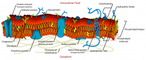
Fig. 2.7. Cell membranes are made of a phospholipid bilayer with embedded proteins.┬ĀThis diagram shows the phospholipid hydrophobic tails pointed toward each other and the hydrophilic heads pointed outward.
The cell membrane is composed of a double layer of phospholipids (Fig. 2.7). Phospholipids are organic molecules that have a non-polar hydrophobic (water-avoiding) end and a polar hydrophilic (water-attracting) end. For more information about non-polar and polar bonds, please see the topic, Types of Covalent Bonds.
┬Ā
These two layers of phospholipids form a bilayer ŌĆ£sandwich.ŌĆØ The hydrophobic ends face each other, forming the sandwich ŌĆ£filling.ŌĆØ The hydrophilic ends form the sandwich ŌĆ£breadŌĆØ on each side of the membrane. Hydrophilic ends thus have contact with the inside and outside of the cell.
┬Ā
The phospholipid cell membrane also has proteins embedded in it. The proteins have various functions. Some proteins act as enzymes to catalyze biological reactions, and other proteins act as channels for transporting molecules in and out of the cell (Fig. 2.7).
┬Ā
┬Ā
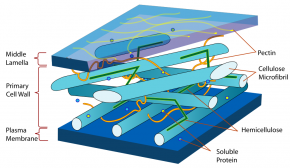
Fig. 2.8. Diagram of a plant cell wall
Cell walls surround the cell membrane and provide protection and structural stability for the organism (Fig. 2.8). Although the cell wall can be rigid, it is usually flexible and allows the diffusion of water and nutrients. Cell walls are present in the cells of eukaryotic plants, algae, and fungi. They are also found in many prokaryotic bacteria, and some archaeabacteria. Animal cells do not have cell walls.
┬Ā
┬Ā
┬Ā
Cellular Water Balance
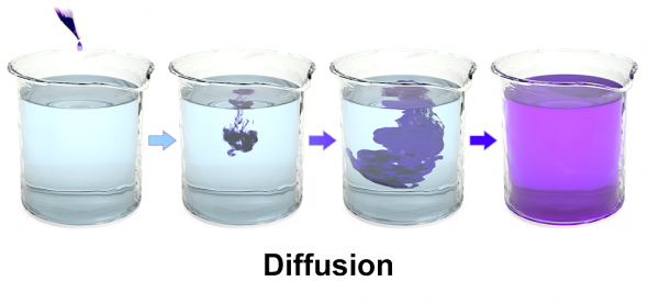
Cell membranes allow a cell to regulate its internal environment. Water and other molecules pass across the cell membrane through diffusion. Diffusion is the tendency for molecules of any substance to spread out into the available space. Diffusion is driven by thermal energy, which causes movement through random vibration of molecules (Fig. 2.9).
┬Ā
Image caption
Fig. 2.9. Concentration gradient leading to diffusion and dynamic equilibrium
Image copyright and source
┬Ā
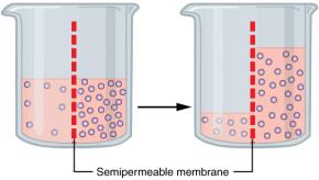
Fig. 2.10. Diffusion across a permeable barrier
Molecules generally travel from areas of high concentration to areas of low concentration and across permeable or semi-permeable barriers (Fig. 2.10). The cell membrane is selectively permeable to molecules of a certain size and charge, meaning some substances pass through the lipid bilayer freely, whereas others must be transported.
┬Ā
Non-polar molecules like carbon dioxide (CO2) and oxygen gas (O2) pass through the cell membrane easily through diffusion. Water (H2O) can also easily cross the membrane. Even though it is a polar molecule, the small size of a water molecule allows it to diffuse across the membrane.
┬Ā
On the other hand, large polar molecules like glucose have big hydrophobic sections. The large size prevents them from passively diffusing across the membrane. In order to pass through, large, polar molecules must be actively transported. Active transport uses energy stored by the cell and is accomplished by proteins embedded in the cell membrane.
┬Ā
The concentration of dissolved substances is often very different inside a cell compared with the outside environment. For example, the inside of a cell tends to be higher in proteins than the outside. The inside of cells also contains higher concentrations of nutrients and dissolved gases necessary for respiration, growth, and reproduction.
┬Ā
The external environment of a cell also contains solutes. Solutes are substances dissolved in fluids, like table salt, which dissolves in water as sodium and chloride. A solution is a fluid that contains dissolved solutes.
┬Ā
┬Ā
Fig. 2.11. The process of osmosis causes water to move from areas of low solute concentration to areas of high solute concentration.
Consider a cell placed in seawater, which contains several dissolved salts and other trace elements. The difference in relative concentration of dissolved substances inside and outside the cell has very important consequences for cell size. Water will diffuse across a membrane from an area of low solute concentration to an area of high solute concentration through a process called osmosis. Osmosis is the special term given to the diffusion of water across a permeable surface. Water will move until the solute concentration of on both sides of the membrane is equal (Fig. 2.11).
┬Ā
Cells deal with the problem of cellular water balance in different ways. Some cells remain at the same concentration as the surrounding environment. In fact, the cells of many marine organisms have very similar solute concentrations as seawater.
┬Ā
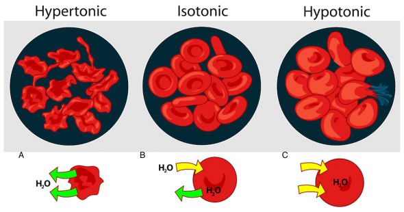
Consider a human blood cell, a type of eukaryotic cell without a cell wall. Blood cells live in an environment with a similar solute concentration as that on the inside of the cell. The environment is said to be isotonic (prefix iso- meaning same) compared to the cell (Fig. 2.12 B). Water will move across the cell membrane in both directions equally, so there is no net movement of water.
┬Ā
Next consider a blood cell that is placed in a hypertonic solution (prefix hyper- meaning more), with a higher concentration of solutes compared to the interior of the cell. Through osmosis, water will move out of the cell, and the cell will shrink (Fig. 2.12 A).
┬Ā
On the other hand, a blood cell placed in a hypotonic (prefix hypo- meaning less) solution with a lower concentration of solutes compared to the cell, will increase in size and be in danger of rupturing and dying (Fig. 2.12 C).
┬Ā
Image caption
Fig. 2.12. Water moves out of (green arrows) or into (yellow arrows) a blood cell, depending on the concentration of solutes in the environment: (A) hypertonic, (B) isotonic, and (C) hypotonic solutions.
Image copyright and source
┬Ā
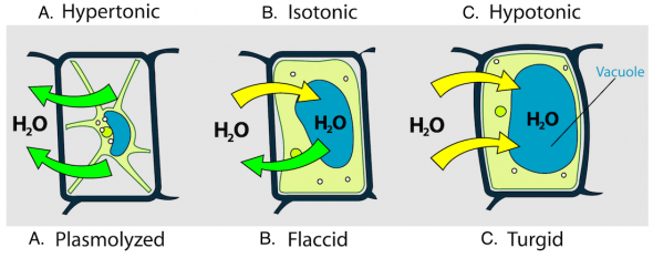
In contrast to animal cells, algae and plant cells have cell walls. A hypotonic solution can cause the cell to become very firm or ŌĆ£turgidŌĆØ because the rigid cell wall pushes back as water flows into the cells. This keeps the entire plant erect, allowing for tall plant stems and upward growth. The rigid structure of the cell wall actually prevents over-expansion (Fig. 2.13 C).
┬Ā
Plant cells placed into isotonic solutions wilt (Fig. 2.13 B). Even though the solute concentrations are equal, plants wilt and become flaccid, without the turgor pressure of the cell wall to stay firm.
┬Ā
In the case of a hypertonic solution, water leaves the cell. The cell membrane will pull away from the cell wall (Fig. 2.13 A). Plasmolysis┬Āis the term used to describe the process of cells┬Ālosing water in a hypertonic solution.
┬Ā
Image caption
Fig. 2.13. Diagram showing plant cell walls (black line), cell membranes (green lines), and relative amount of water (blue ovals) inside the cell. The arrows show the direction of water moving in (yellow) and out (green) of a plant cell placed in these environmental conditions: (A) hypertonic, (B) isotonic, and (C) hypotonic solutions. The movement of water into or out of these plant cells cause (A) plasmolyzed (shrunken), (B) flaccid (wilted), and (C) turgid (firm) cell conditions.
Image copyright and source
┬Ā
Osmosis, as described above in blood and plant cells, is a passive movement of water. It is important to note that some organisms also actively transport water into or out of the cell. This process is called osmoregulation, and it requires energy input by the organism.
┬Ā
Cellular Components
┬Ā
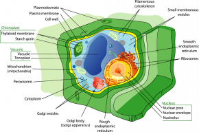
Fig. 2.14. Diagram of a eukaryotic plant cell
Organelles are specialized subunits within the cell. Organelles are usually enclosed in another lipid membrane. The nucleus is an organelle found in eukaryotes that contains the genetic material of the cell and controls cell processes. The interior cell structures are surrounded by cytoplasm.┬ĀCytoplasm is a gel like substance made mostly of water.
Organelles compartmentalize cell tasks. Eukaryotes have several important organelles (Fig. 2.14). For example,
- mitochondria are known as the ŌĆ£cellular power plantsŌĆØ because they supply much of the cellŌĆÖs chemical energy.
- ribosomes and the rough endoplasmic reticulum are organelles responsible for protein synthesis.
- smooth endoplasmic reticulum is responsible for metabolism and detoxification.
- golgi apparatus is composed of stacks of membranes and is responsible for packaging and sorting proteins made within the cell.
┬Ā
Eukaryotic cells contain a membrane-bound nucleus and other organelles. Their cells are range in size from 10 to 100 micrometers (╬╝m). Eukaryotic organisms can be unicellular or multicellular. The nucleus of eukaryotic cells contains at least┬Ātwo chromosomes. A chromosome is a single piece of DNA that contains many genes.
┬Ā
Prokaryotes, on the other hand, have much smaller cells (1ŌĆō10 ┬Ąm) and are only unicellular. Prokaryotes do not have a nucleus or any other membrane-bound organelles. In prokaryotes, their DNA is contained within a single, circular chromosome inside the cytoplasm.
┬Ā
┬Ā
┬Ā
Cells of Photosynthesizers
All photosynthetic autotrophs have structures inside their cells that harvest and convert sunlight into usable energy. However, there are differences between prokaryotic and eukaryotic cells in terms of where photosynthesis occurs.
┬Ā
Prokaryotic autotrophs perform photosynthesis and respiration within thylakoid membranes. Thylakoid membranes are generally located near the outer cell membrane and contain pigments like chlorophyll that are responsible for photosynthesis.
┬Ā
┬Ā
Fig. 2.15. Diagram of a chloroplast
Eukaryotic autotrophs are more complex. Most eukaryotic autotrophs have organelles called chloroplasts that contain the machinery for photosynthesis. The inside of the chloroplast is filled with stroma, a watery substance similar to cytoplasm. Thylakoids containing the photosynthetic pigments are located within the stroma (Fig. 2.15). Grana are arranged stacks of disc-shaped thylakoids.
┬Ā
Interestingly, cyanobacteria are considered the ancestors of modern chloroplasts. Scientists think cyanobacteria were incorporated into eukaryotic cells through a symbiotic relationship. A free-living cyanobacteria was likely taken inside the eukaryotic cell as a helpful organism and eventually became part of the host. This same process also likely occurred for eukaryotic mitochondria.
┬Ā
There is evidence for chloroplasts and mitochondria previously being separate organisms:
- Both contain their own DNA. The DNA in chloroplasts and mitochondria is separate from the DNA in the cellŌĆÖs nucleus.
┬Ā
- The chloroplast and mitochondria organelles also have two membranes. The second membrane is thought to have been produced when the hostŌĆÖs cell membrane surrounded a cyanobacterium as it entered the host cell.
┬Ā
┬Ā
Activity
Observe the cell membrane, cell wall, and chloroplasts of an aquatic plant.
┬Ā
Plant Structure
Plants are photosynthetic organisms. Almost all plants have vein-like vascular tissue and structural systems that transport water and nutrients throughout the plant. There are some plants without vascular tissuesŌĆömoss-like species that roughly resemble some seaweeds or algae. This section describes vein-like vascular plants.
┬Ā
┬Ā
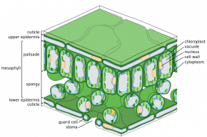
Fig. 2.16. Anatomy of a typical vascular plant leaf
The body plan of plants is generally broken down into three systems: the leaves, the root, and the shoot. The leaves of the plant are the primary location of photosynthesis (Fig. 2.16). Roots provide an anchoring system for the plant, in addition to absorption. For most plants, water and nutrients are absorbed through the roots. The shoot system provides a method of exchanging water and nutrients between the roots and the leaves through a network of vascular tissue. The shoot system also allows plants to grow vertically. Sunlight can be a limiting resource if the plant is shaded, so having the ability to grow taller than plant neighbors is an important adaptation for plants.
┬Ā
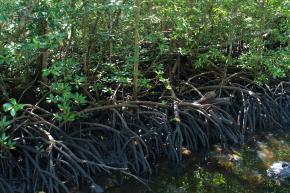
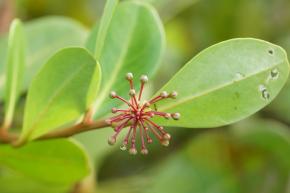
Although there is a great diversity of plants that live on land, there are far fewer plants that live in aquatic habitats. Examples of aquatic plants include seagrasses and mangroves (Fig. 2.17).
┬Ā
Fig. 2.17. (A) Mangrove roots absorb water and nutrients and store starch reserves.
Fig. 2.17. (B) Mangrove shoots transport water and nutrients.
Microalgae
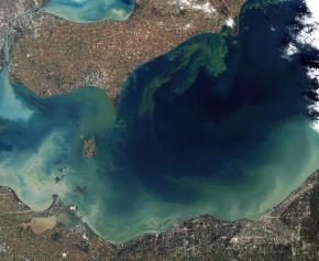
Microalgae are single-celled, photosynthetic autotrophs that live in the water. Although individual cells are too small to be visible to the naked eye, microalgae can be seen as colorful plumes (Fig. 2.18 A). Microalgae have amazing diversity in terms of their structure and function.
┬Ā
┬Ā
Fig. 2.18. (A) A satellite image of an algae bloom in Lake Erie shows swirls of phytoplankton microalgae.
Image courtesy of National Aeronautics and Space Administration (NASA)
Fig. 2.18.┬Ā(B) A chain of several individual phytoplankton microalgae cells under a light microscope
Image courtesy of National Oceanic and Atmospheric Administration (NOAA)
┬Ā
Phytoplankton are microalgae that float in the water. Phytoplankton produce approximately half of EarthŌĆÖs oxygen via photosynthesis and also provide food for many aquatic organisms. It is important for photosynthetic organisms to be near sunlight, thus microalgae must remain near the oceanŌĆÖs surface. As a result, many phytoplankton have evolved structural adaptations that prevent sinking, such as oil-filled sacs or spines (Fig. 2.18 B). Other microalgae, like cyanobacteria, cover the seafloor in shallow areas where they have access to light.
┬Ā
For more information about microalgae, please see the topic, Marine Microbes.
┬Ā
Macroalgae
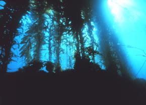
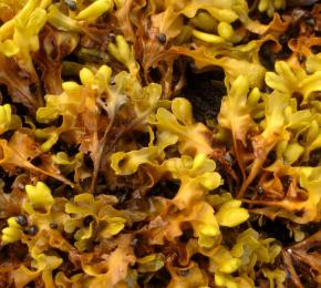
Macroalgae are algae visible to the naked eye. Macroalgae are sometimes referred to as ŌĆ£seaweed,ŌĆØ and they have a wide array of body plans and structures. Macroalgae can be as small as a few millimeters or as large as 45 meters, as in the case of giant kelp (Macrocystis pyrifera, Fig. 2.19). See Table 2.3 for common algae terminology.
┬Ā
Fig. 2.19. (A) Petticoat algae, Padina pavonica, a phaeophyte or brown macroalga
Fig. 2.19.┬Ā(B) Giant kelp, Macrocystis pyrifera, a phaeophyte or brown macroalga
Image courtesy of National Oceanic and Atmospheric Administration (NOAA)
Table 2.3. Common algae terms
| Blade ŌĆō a large, flattened branch of the thallus (Fig. 2.20) |
| Calcified ŌĆō describes an organism with calcium carbonate in its tissue, which often gives it a hard texture |
| Calcium carbonate ŌĆō a chemical compound found in some organisms for structure and defense |
| Dichotomous ŌĆō a branching pattern where branches are divided in two (Fig. 2.24 C) |
| Filaments or filamentous ŌĆōvery thin branches that are usually chains of connected single cells (Fig. 2.24 B) |
| Holdfast ŌĆō part of the thallus that anchors the plant to the bottom (rock, sand, coral rubble, other plants, or animals; Fig. 2.20) |
| Midrib ŌĆō a thickened portion in the center of an alga blade or stipe. The mid rib may look similar to a vein, however it is not true vascular tissue (Fig. 2.21 A). |
| Pneumatocyst ŌĆō air bladders used to hold the alga upright in the water (Fig. 2.22 A). |
| Segmented ŌĆō visibly divided into separate uniform parts or sections (Fig. 2.22 A). |
| Stipe ŌĆō a stem-like structure. Unlike terrestrial plants, the stipe does not have true vascular tissue (Fig. 2.20). |
| Thallus ŌĆō main body of the alga |
┬Ā
Since algae lack vascular tissue, they do not have true shoots, stems, leaves, or roots. A thallus is the term used to describe the main body of an alga (Fig. 2.20).
┬Ā
Fig. 2.20. (A) Diagram showing general alga morphology
Fig. 2.20. (B) Giant kelp, Macrocystis pyrifera with the holdfast, stipe, float, and blades visible.
┬Ā
Blades are the flattened portions of the thallus. Algae blades come in a variety of shapes and sizes (Fig 2.21). Some blades even have a thickened midrib very similar in appearance to plant leaves.
┬Ā
Fig. 2.21. (A) Toothed wrack seaweed, Fucus serratus, blades with thickened midribs
Fig. 2.21.┬Ā(B) Giant Kelp, Macrocystis pyrifera, blades
Image courtesy of National Oceanic and Atmospheric Administration (NOAA)
┬Ā
Fig. 2.21. (C) MermaidŌĆÖs fan algae, Udotea flabellum
Image courtesy of James St. John, Flickr
Fig. 2.21.┬Ā(D) Antarctic Kelp, Durvillaea Antarctica, blades
┬Ā
Some macroalgae also have a stipe, which is a stem-like portion of the thallus (Fig. 2.22).
┬Ā
Fig. 2.22. (A) Loops of stem-like stipes with pneumatocysts from Macrocystis pyrifera
Image courtesy of jariceiii, Flickr
Fig. 2.22. (B) Stipes on this red algae support small blades
Image courtesy of Ed Bierman, Flickr
┬Ā
Fig. 2.22. (C) A leafy algae with short stipes
Image courtesy of toothbrush party, Flickr
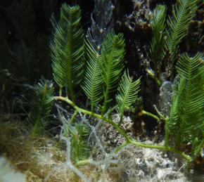
Fig. 2.22. (D)┬ĀLong stipes of Caulerpa sertularioides with feather-like blades attached
┬Ā
The holdfast anchors macroalgae to the substrate. This is very important, especially in areas with high currents or wave action. When an alga is broken off above the holdfast, the holdfast may also re-grow into a new alga.
┬Ā
Holdfasts are similar to plant roots in that both holdfasts and roots anchor the organism in place. But unlike holdfasts, plant roots are specialized to store food and absorb nutrients for transport throughout the plant.
┬Ā
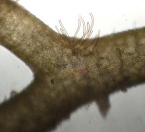
Fig. 2.23. (A) Alga holdfast under the microscope
Fig. 2.23.┬Ā(B) Algae Holdfast attached to rock
┬Ā
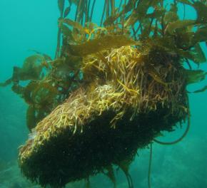
Fig. 2.23.┬Ā(C) Giant kelp holdfast
Image courtesy of Claire Fackler, National Oceanic and Atmospheric Administration (NOAA)
Fig. 2.23.┬Ā(D) Holdfasts attached to pebble substrate
Image courtesy of Nadya Peek, Flickr
┬Ā
┬Ā
Fig. 2.24. (A) Example of a segmented thallus, (B) Example of a filamentous thallus, and (C) Example of a dichotomous thallus.
Image courtesy of Cataneo et Fernandes; Delle Chiaie, S.; Pietro, Marc di, Wikimedia Commons;┬ĀDiagrams by Narrissa Spies
Body plans of macroalgae range widely. In some macroalgae, the alga thallus has distinct parts. For example, the thallus can be divided into branches. Some algae have┬ĀBody plans of macroalgae range widely. In some macroalgae, the alga thallus has distinct parts. For example, the thallus can be divided into branches. Some algae have Dichotomous branching, where every branch node divides into two new branches. Fig. 2.24 C shows Dictyota sp., a species of brown alga, with dichotomous branching.
┬Ā
A segmented thallus occurs when an alga is divided into distinct sections. Halimeda opuntia is a green alga with segmented branches (Fig. 2.24 A). In some algae, like the genus Griffithsia, long chains of individual cells form filaments (Fig. 2.24 B).
┬Ā
┬Ā
Activity
Identify parts of algae and compare algae to vascular plants.
